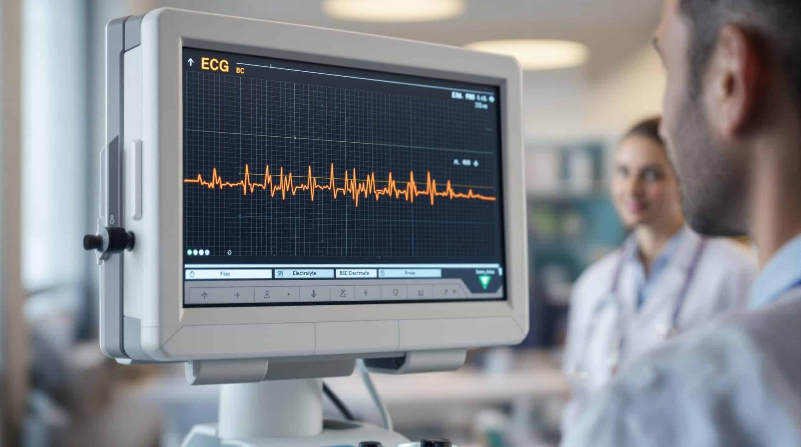
Electrolyte imbalances can cause noticeable changes in ECG readings, which help in early diagnosis and treatment of cardiac risks. Here’s a quick summary of key imbalances and their ECG effects:
- Hyperkalemia: Peaked T waves, widened QRS, sine wave pattern in severe cases.
- Hypokalemia: Flattened T waves, ST depression, prominent U waves.
- Hypercalcemia: Shortened QT interval.
- Hypocalcemia: Prolonged QT interval.
- Hypermagnesemia: Bradycardia, prolonged QT.
- Hypomagnesemia: QT prolongation, T-wave changes.
Why It Matters:
- Early ECG changes can prevent complications like arrhythmias.
- Continuous monitoring is crucial, especially in critical care.
- Treatments like IV potassium or calcium depend on specific ECG findings.
| Electrolyte Imbalance | Early ECG Signs | Severe ECG Changes |
|---|---|---|
| Hyperkalemia | Peaked T waves | Sine wave pattern |
| Hypokalemia | T wave flattening | Prominent U waves |
| Hypercalcemia | Shortened QT interval | Bradycardia |
| Hypocalcemia | Prolonged QT interval | T wave inversion |
Understanding these patterns is essential for timely interventions and better patient outcomes.
Electrolyte Imbalances and Their ECG Effects
Hyperkalemia and ECG Changes
Hyperkalemia causes noticeable ECG changes that worsen as potassium levels increase. One of the earliest signs is the development of tall, peaked T waves. As potassium levels rise further, P waves may flatten or disappear, and the QRS complex becomes significantly wider. In extreme cases, the ECG can show a sine wave pattern, a critical sign requiring immediate intervention.
While hyperkalemia has its own distinct effects, low potassium levels (hypokalemia) present a different set of challenges on the ECG.
Hypokalemia and ECG Changes
When potassium levels are low, the ECG shows specific abnormalities that aid in diagnosis and treatment. Common features include flattened or inverted T waves, ST segment depression, prominent U waves, and a longer P wave duration.
| ECG Feature | Changes in Hypokalemia |
|---|---|
| T waves | Flattened or inverted |
| ST segment | Depression |
| U waves | Prominent, especially in precordial leads |
| P wave | Increased duration |
These changes become more noticeable as potassium levels drop further, making ECG monitoring a crucial tool for evaluating the severity of hypokalemia and guiding treatment decisions [3].
Calcium Imbalances and ECG Changes
Calcium levels also play a key role in cardiac conduction. Hypercalcemia typically shortens the QT interval, while hypocalcemia prolongs it. These changes are important diagnostic clues and can help predict the likelihood of dangerous arrhythmias.
Magnesium levels, often linked with calcium and potassium imbalances, also have a significant impact on ECG readings.
Magnesium Imbalances and ECG Changes
Magnesium disturbances often occur alongside potassium and calcium issues, making it essential to monitor all electrolytes together. Hypermagnesemia is associated with bradycardia and a prolonged QT interval. On the other hand, hypomagnesemia can cause QT prolongation and noticeable T-wave changes, increasing the risk of severe arrhythmias.
Interestingly, the degree of ECG changes doesn’t always match the severity of the electrolyte imbalance [2]. Even mild disturbances can cause pronounced ECG shifts, especially in patients with pre-existing heart conditions. This unpredictability underscores the need for continuous ECG monitoring and frequent checks of electrolyte levels to ensure accurate assessment and timely treatment.
Understanding EKG Changes Due to Electrolyte Abnormalities
Using ECGs for Diagnosis and Risk Assessment
Building on the knowledge of how specific ECG changes relate to electrolyte imbalances, this section explains how these insights help with early detection and risk evaluation.
Detecting Electrolyte Imbalances Early
An ECG can act as an early warning tool, identifying electrolyte imbalances before they escalate into life-threatening situations. Despite its importance, about 30% of patients presenting with severe hypokalemia (≤2.5 mEq/L) in emergency departments don’t undergo ECG screening [3].
Changes in ECG readings follow recognizable patterns, giving clinicians a way to assess the severity of electrolyte imbalances. Recognizing these patterns early allows for timely medical action and helps avoid serious health issues.
| Electrolyte Imbalance | Early ECG Signs | Advanced ECG Signs |
|---|---|---|
| Hyperkalemia | Peaked T waves | Flattened P waves, sine wave |
| Hypokalemia | T wave flattening | ST depression, U waves |
| Hypercalcemia | Shortened QT | Bradycardia |
| Hypocalcemia | Prolonged QT | ST changes, T wave inversion |
These warning signs are essential for guiding interventions and preventing complications that could endanger a patient’s life.
Assessing Arrhythmia Risk
Spotting ECG changes early also helps clinicians evaluate and manage arrhythmia risks. The relationship between serum electrolyte levels and ECG changes isn’t always straightforward, so continuous monitoring and a comprehensive approach are necessary [6].
"Studies have shown that ECG changes can predict clinical outcomes such as arrhythmias and cardiac arrest. For example, a study found that 77% of patients with severe hypokalemia who had ECG abnormalities required IV potassium replacement, underscoring the importance of ECG in identifying high-risk patients" [3].
Widened QRS complexes or ST changes often indicate a high likelihood of arrhythmias, necessitating urgent care [2][4]. Clinicians must stay alert, as ECG changes linked to electrolyte imbalances can sometimes resemble other conditions, such as ischemia, making accurate diagnosis essential [1][3].
Key practices for monitoring include:
- Continuous ECG observation for patients with known electrolyte imbalances
- Evaluating multiple factors when determining arrhythmia risk
- Acting promptly when concerning ECG changes are detected
sbb-itb-aa73634
Managing Electrolyte Imbalances with ECG Monitoring
Treatments Based on ECG Findings
Clinicians rely on ECG abnormalities to decide on the best treatments and to monitor how well those treatments are working. Once specific ECG changes are identified during diagnosis, they help shape targeted interventions based on the patient’s condition.
For hyperkalemia, the treatment depends on how the ECG changes evolve:
| ECG Finding | Treatment Approach | Monitoring Focus |
|---|---|---|
| Peaked T waves | Insulin with glucose, beta-2 agonists | Watch for T wave changes |
| Widened QRS | Immediate calcium administration | Monitor QRS widening |
| Sine wave pattern | Emergency measures like dialysis | Track cardiac rhythm |
Treatment plans must align with the type and severity of the electrolyte imbalance. For example, severe hypokalemia, often indicated by U-waves and ST segment depression, calls for aggressive potassium replacement therapy [1].
"ECG can be a predictive tool to diagnose severe hyperkalemia and recognize which hyperkalemia patients are in danger of adverse episodes." [5]
While these targeted treatments address immediate risks, continuous ECG monitoring plays a key role in ensuring patient safety and guiding any additional interventions.
Continuous ECG Monitoring in Care
Real-time ECG monitoring is a cornerstone of managing electrolyte imbalances, especially in critical care settings. Studies show that 56% of patients with severe hyperkalemia (≥ 8 mmol/L) experience adverse episodes [5].
Specific monitoring protocols in critical care focus on:
| Monitoring Aspect | Purpose | Trigger for Action |
|---|---|---|
| Rhythm analysis | Detect arrhythmias | Immediate intervention |
| ST segment tracking | Spot changes | Adjust treatment |
| T wave morphology | Monitor hyperkalemia signs | Escalate protocols |
Even minor electrolyte imbalances can cause notable ECG changes, particularly in patients with existing heart conditions [2].
Study Tools for Nursing Students
For nursing students gearing up for the NGN NCLEX, understanding ECG interpretation is a key skill, especially when it comes to managing electrolyte imbalances. Today’s educational platforms are tailored to address the challenges of licensure exam preparation.
Nurse Cram NCLEX Exam Review

Nurse Cram provides focused study materials to help nursing students break down the complexities of ECG interpretation, particularly in relation to electrolyte imbalances. Its modules simplify intricate ECG patterns, making them easier to grasp.
The platform incorporates scenarios, case studies, and NGN-style questions to enhance learning:
| Study Component | Learning Focus | Clinical Application |
|---|---|---|
| Scenario-based Exercises | ECG changes linked to electrolytes | Analyzing real patient cases |
| Interactive Case Studies | Recognizing ECG patterns | Handling critical care situations |
| NGN-style Questions | Strengthening clinical judgment | Prioritizing treatments |
Nurse Cram prioritizes building critical thinking skills, enabling students to identify and manage ECG changes caused by electrolyte imbalances. From spotting early warning signs to addressing severe disturbances, students gain practical expertise.
"The platform guides students through effective test-taking strategies and reinforces essential critical thinking and clinical judgment skills, setting the stage for a successful nursing career."
Interactive questions, like bow-tie and matrix grid formats, further sharpen ECG interpretation skills, helping students excel on the NGN NCLEX. By mastering these patterns, students are better prepared for timely interventions, ultimately leading to better patient care. These tools seamlessly connect classroom learning with clinical practice, ensuring students are ready for both exams and real-world nursing challenges.
Conclusion: ECG Changes and Electrolyte Imbalances
Continuous ECG monitoring plays a key role in patient care, especially when dealing with electrolyte imbalances. Missed opportunities in its use underline the need for better clinical practices and early intervention strategies.
ECG changes act as an early warning system, allowing healthcare providers to address issues before complications develop. For example, the Institute for Algorithmic Medicine emphasizes the importance of recognizing these changes early, particularly in conditions like severe hyperkalemia [6][2][4].
Here’s why continuous ECG monitoring makes a difference:
| Monitoring Approach | Clinical Impact |
|---|---|
| Real-time Detection | Enables immediate intervention |
| Regular Assessment | Helps adjust treatments as needed |
Identifying patterns such as flattened T waves or sine waves is critical for timely action [1][2][4]. When combined with a thorough clinical assessment, this approach lays the groundwork for effective care. By integrating consistent monitoring with prompt responses, healthcare providers can better manage electrolyte imbalances.
As advancements in healthcare continue, focusing on ECG interpretation alongside clinical evaluations will remain essential. This combination of vigilant monitoring and swift action ensures improved outcomes for patients dealing with electrolyte disturbances.
FAQs
What electrolyte imbalances cause ECG changes?
Certain electrolyte imbalances can lead to noticeable changes in ECG patterns. Here’s a quick breakdown:
| Electrolyte Imbalance | Common ECG Changes |
|---|---|
| Hyperkalemia | Peaked T waves, PR prolongation, absent P waves |
| Hypokalemia | Flattened T waves, ST depression, U waves |
| Hypercalcemia | Shortened QT interval |
| Hypocalcemia | Prolonged QT interval |
| Hypermagnesemia | Prolonged PR and QT intervals |
| Hypomagnesemia | Widened QRS complex, T wave changes |
It’s worth noting that ECG changes don’t always match the severity of the electrolyte imbalance. Continuous monitoring is especially important for patients with heart conditions [2][7].
What does electrolyte imbalance look like on an ECG?
Electrolyte imbalances often show distinct patterns on an ECG. For example, hyperkalemia can be identified by its progression:
- Early Signs: Peaked T waves
- Intermediate Signs: Prolonged PR interval and flattened P waves
- Late Signs: Widened QRS complex, which may progress to a sine wave pattern [2][7]
On the other hand, hypokalemia is marked by T-wave flattening, ST depression, and the appearance of prominent U-waves, which become more pronounced as the imbalance worsens [1][3].
For hyperkalemia, administering IV calcium can quickly reverse ECG changes, though it doesn’t lower potassium levels [8].
Recognizing these patterns is crucial for effective diagnosis and treatment. These insights are also valuable for NGN NCLEX preparation, as noted in resources like Nurse Cram, helping guide timely interventions and improve patient outcomes.
Related posts
- Top 20 NCLEX Pharmacology Questions and Answers
- PQRST Wave Basics for ECG Interpretation
- ECG Waveform Components: Quick Reference Guide
- How to Use ABCDE for Patient Deterioration

Mia is dedicated to helping nursing students and new graduates confidently prepare for the Next Generation NCLEX exam. With a focus on providing clear, actionable advice and support, Mia offers practical study tips, effective strategies, and encouragement to guide you through the complexities of nursing exams. Whether you need help mastering question formats, managing stress, or creating a personalized study plan, Mia is here to ensure you feel prepared and empowered every step of the way.

